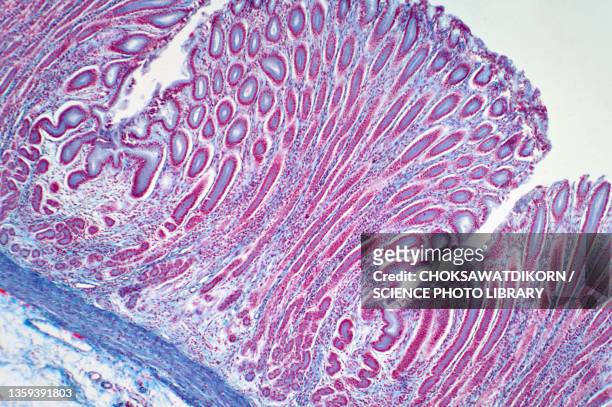
hair follicle, hair bulb, and hair papilla, human scalp, 25x at 35mm. growth takes place at the base of the hair follicle. adipose tissue surrounds the hair bulb. - lichtmicroscopische foto stockfoto's en -beelden
alfalfa (medicago sativa) root nodosity cross section - lichtmicroscopische foto stockfoto's en -beelden
onion root cells undergoing mitosis, seen under an optical microscope - lichtmicroscopische foto stockfoto's en -beelden
microscopische mening van dwarsdoorsnede van de wortel van de varen - lichtmicroscopische foto stockfoto's en -beelden
stem cross section. buttercup (ranunculus), herbaceous dicot, 8x. shows: vascular bundles arranged in a ring (typical of dicots), xylem, phloem, epidermis, cortex, and pith. - lichtmicroscopische foto stockfoto's en -beelden
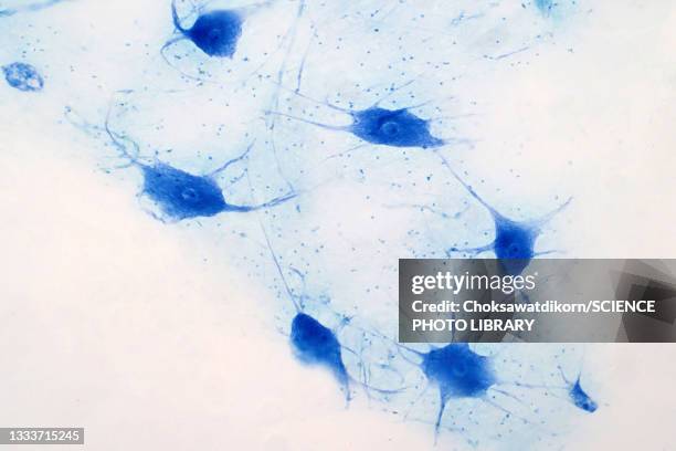
immunofluorescent photomicrograph, organs samples, histological examination, histopathology on the microscope - lichtmicroscopische foto stockfoto's en -beelden
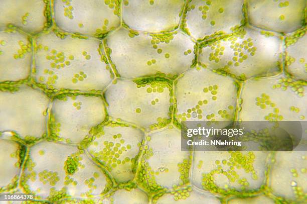
microscopische weergave van de dwarsdoorsnede van lila blad - lichtmicroscopische foto stockfoto's en -beelden
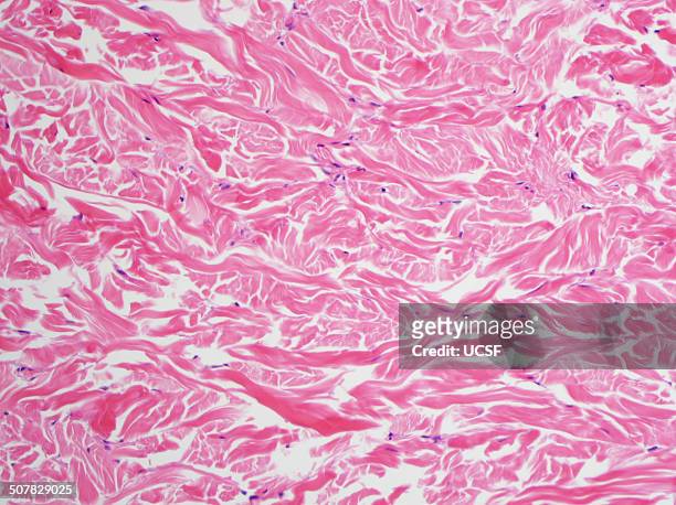
h&e stain, light microscopy, abundant collagen in a gardner fibroma - lichtmicroscopische foto stockfoto's en -beelden
cheek cell. human squamous epithelial cell, mouth, 250x. shows: nucleus, cytoplastm and cell membrane. this is a very flat (or squamous) cell obtained inside the oral cavity. iodine stain. - lichtmicroscopische foto stockfoto's en -beelden
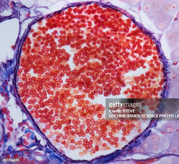
atherosclerosis of coronary artery, 5x at 35mm. reduced lumen. wall has excess calcification & fibrous connective tissue. h - lichtmicroscopische foto stockfoto's en -beelden
leaf cross section, lilac (syringa), 100x. shows: palisade mesophyll, spongy mesophyll, stoma, guard cells, epidermis, and a small vein that shows xylem and phloem. - lichtmicroscopische foto stockfoto's en -beelden
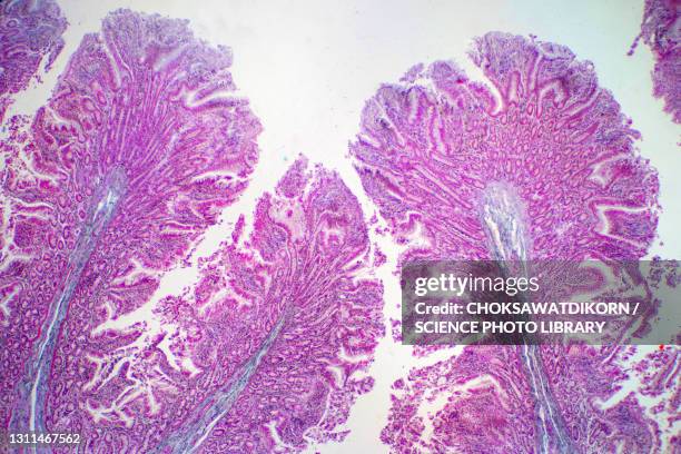
skin. desquamation (sloughing of cells) from the epidermis, thick skin, human, 100x at 35mm. shows: epidermal layers (stratum: corneum, granulosum, spinosum, basale), dermis, sweat gland ducts and desquamating cells sloughing off the surface. - lichtmicroscopische foto stockfoto's en -beelden
smooth muscle longitudinal section,100x light micrograph - lichtmicroscopische foto stockfoto's en -beelden
Photomicrograph of the malaria parasite Plasmodium Ovale growing as a double trophozoite in one red blood cell and a single trophozoite, on a thin...
Aluminium 4% copper . Aluminium 4% copper . Grains of solid solution of copper in aluminium. Light micrograph in bright field. Magnification 500x.
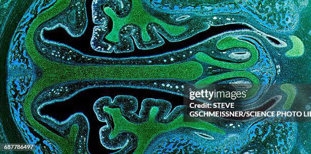
Carbon fibre in a polymer matrix. PMC carbon fibre in a polymer matrix. Light micrograph in crossed polar light. Magnification 100x.
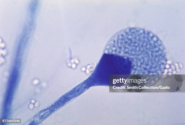
Light micrograph of mature sporangium of a Mucor fungus, 1955. Image courtesy CDC/Dr Lucille K Georg. Image courtesy CDC.
Micrograph revealing histologic changes in human skin infected with the smallpox variola virus, 1975. Image courtesy CDC. .
Micrograph showing the histologic changes in human skin infected with smallpox, prepared with hematoxylin-eosin stain, 1975. .
Magnified 1125X, this thin-film, Giemsa-stained photomicrograph revealed the presence of a growing Plasmodium ovale trophozoite, with a ring nucleus....
histology sample tactile corpuscle sec. under light microscopy with white background - lichtmicroscopische foto stockfoto's en -beelden